 Theory of Evolution Cartoonishly
Dumb
Part Three:
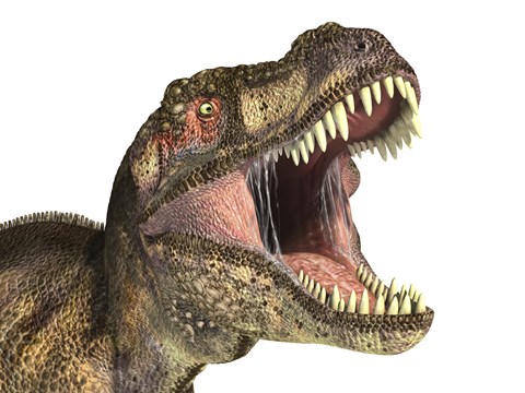
Very Young Tyrannosaurus Rex Dinosaur
“Researchers working on ancient DNA had claimed
previously that they had recovered DNA millions of years old, but subsequent
work failed to validate
the results. The only widely accepted claims of
ancient molecules were
no more than several tens of thousands of years old”.
Mary H. Schweitzer
Part One of this series:
re-visited a series of letters by Polish
professor Maciej Giertych exposing evolution:
The theory of evolution is maintained for ideological
reasons and not because scientific evidence supports it. If it were not for the
lack of another atheistic explanation of the origin of life and of all its
forms, this theory would have been dismissed by scientists long ago. In fact most
scientists prefer not to get involved in the controversy over evolution because
of the possible consequences to their careers. A recent example of such consequences for Dr.
Rick Von Sternberg of the Smithsonian Institution can be seen discussed in a National
Review article[1]. Most biologists can work in their own fields and
advance academically without ever mentioning evolution and most choose not
to mention it. ….
Part
Two:
recalled, amongst other things, that classic quote about evolution by
G. K. Chesterton:
“The evolutionists seem to know everything
about the missing link
except the fact that it is missing”.
Here
now, in Part Three, is another
telling piece of evidence:
Evolution Falsified, Once Again
Evolution
Tuesday, 02
August 2011 11:26
R. Sungenis:
In this article, field researcher Mary H. Schweitzer writes in the most
prestigious science magazine today, Scientific American, about her
discovery of soft tissue and blood cells in the bone of a Tyrannosaurus rex
dinosaur that, according to modern evolutionary dating techniques, is about 70
million years old. If it hasn’t struck you already, science tells us that
organic tissue could barely last 7,000 years, much less 10,000 times 7,000
years. So what does science do with this anomaly? It pleads ignorance, and it
does so while it tries to find a way to dismiss the evidence. When Ms.
Schweitzer brought her evidence to Jack Horner, curator of paleontology at the museum and one of
the world’s foremost dinosaur authorities, after a long look under the
microscope at the nucleated blood cells of the T-Rex, he said to Ms.
Schweitzer: “So prove to me they aren’t.” That about sums up the history of the
bias and deliberate attempts to twist the evidence in favor of evolution that
occurs on a daily basis in our high school and college classrooms. Whereas Ms.
Schweitzer’s find should have been hailed as one of the most astounding
discoveries in history since Darwin wrote his book on the evolutionary
hypothesis in 1879, she is basically assigned the impossible task of finding a
way to dismiss the blood cell’s prima facie denial of evolution, and
implied in that “request” is the fact that she will lose her job if she doesn’t
seek an alternative answer. What does Ms. Schweitzer decide to do? The next
sentence in her story tells us loud and clear. She capitulates to the reigning
paradigm of modern science, without question: “It was an irresistible
challenge, and one that has helped frame how I ask my research questions, even
now.” So Ms. Schweitzer, in order to continue to be a member of the status quo
and receive her pay check from the powers-that-be, remains an ardent
evolutionist, seeking to deny the common sense knowledge her heart and mind
scream at her about what it means to see blood cells in dinosaur remains.
“Blood From Stone”
By Mary H. Schweitzer
From Scientific American, December 2010
Peering through the microscope at the thin
slice of fossilized bone, I stared in disbelief at the small red spheres a
colleague had just pointed out to me. The tiny structures lay in a blood vessel
channel that wound through the pale yellow hard tissue. Each had a dark center
resembling a cell nucleus. In fact, the spheres looked just like the blood
cells in reptiles, birds and all other vertebrates alive today except mammals,
whose circulating blood cells lack a nucleus. They couldn’t be cells, I told
myself. The bone slice was from a dinosaur that a team from the Museum of the
Rockies in Bozeman, Mont., had recently uncovered a Tyrannosaurus rex
that died some 67 million years ago--and everyone knew organic material was far
too delicate to persist for such a vast stretch of time.
For more than 300 years paleontologists
have operated under the assumption that the information contained in fossilized
bones lies strictly in the size and shape of the bones themselves. The
conventional wisdom holds that when an animal dies under conditions suitable
for fossilization, inert minerals from the surrounding environment eventually
replace all of the organic molecules—such as those that make up cells, tissues,
pigments and proteins—leaving behind bones composed entirely of mineral. As I
sat in the museum that afternoon in 1992, staring at the crimson structures in
the dinosaur bone, I was actually looking at a sign that this bedrock tenet of
paleontology might not always be true—though at the time, I was mostly puzzled.
Given that dinosaurs were nonmammalian vertebrates, they would have had
nucleated blood cells, and the red items certainly looked the part, but so,
too, they could have arisen from some geologic process unfamiliar to me.
Back then I was a relatively new graduate
student at Montana State University, studying the microstructure of dinosaur
bone, hardly a seasoned pro. After I sought opinions on the identity of the red
spheres from faculty members and other graduate students, word of the puzzle
reached Jack Horner, curator
of paleontology at the museum and one of the world’s foremost dinosaur
authorities. He took a look for himself. Brows furrowed, he gazed through the
microscope for what seemed like hours without saying a word. Then, looking up
at me with a frown, he asked, “What do you think they are?” I replied that I did not know, but they were
the right size, shape and color to be blood cells, and they were in the right
place, too. He grunted, “So prove to me they aren’t.” It was an irresistible
challenge, and one that has helped frame how I ask my research questions, even
now.
Since then, my colleagues and I have
recovered various types of organic remains—including blood vessels, bone cells
and bits of the fingernail-like material that makes up claws—from multiple
specimens, indicating that although soft-tissue preservation in fossils may not
be common, neither is it a one-time occurrence. These findings not only diverge
from textbook description of the fossilization process, they are also yielding
fresh insights into the biology of bygone creatures. For instance, bone from
another T.rex specimen has revealed that the animal was a female that
was “in lay” (preparing to lay eggs) when she died—information we could not have
gleaned from the shape and size of the bones alone. And a protein detected in
remnants of fibers near a small carnivorous dinosaur unearthed in Mongolia has
helped establish that the dinosaur had feathers that, at the molecular level,
resembled those of birds.
Our results have met with a lot of
skepticism—they are, after all, extremely surprising. But the skepticism is a
proper part of science, and I continue to find the work fascinating and full of
promise. The study of ancient organic molecules from dinosaurs has the
potential to advance understanding of the evolution and extinction of these
magnificent creatures in ways we could not have imagined just two decades ago.
FIRST SIGNS
Extraordinary claims, as the old adage
goes, require extraordinary evidence. Careful scientists make every effort to
disprove cherished hypotheses before they accept that their ideas are correct.
Thus, for the past 20 years I have been trying every experiment I can think of
to disprove the hypothesis that the materials my collaborators and I have
discovered are components of soft tissues from dinosaurs and other long-gone
animals.
In the case of the red microstructures
saw in the T.rex bone, I started by thinking that if they were related
to blood cells or to blood cell constituents (such as molecules of hemoglobin
or heme that had clumped together after being released from dying blood cells),
they would have persisted in some, albeit possibly very altered, form only if
the bones themselves were exceptionally well preserved. Such tissue would have
disappeared in poorly preserved skeletons. At the macroscopic level, this was
clearly true. The skeleton, a nearly complete specimen from eastern
Montana—officially named MOR 555 and affectionately dubbed “Big Mike”—includes
many rarely preserved bones. Microscope examination of thin sections of the
limb bones revealed similarly pristine preservation. Most of the blood vessel
channels in the dense bone were empty, not filled with mineral deposits as is
usually the case with dinosaurs. And those ruby microscopic structures appeared
only in the vessel channel, never in the surrounding bone or in sediments
adjacent to the bones, just as should be true of blood cells.
Next, I turned my attention to the
chemical composition of the blood cell look-alikes. Analyses showed that they
were rich in iron, as red blood cells are, and that the iron was specific to
them. Not only did the elemental makeup of the mysterious red things (we
nicknamed them LLRTs, “little round red things”) differ from that of the bone
immediately surrounding the vessel channels, it was also utterly distinct from
that of the sediments in which the dinosaur was buried. But to further test the
connection between the red structures and blood cells, I wanted to examine my
samples for heme, the small iron-containing molecule that gives vertebrate
blood its scarlet hue and enables hemoglobin proteins to carry oxygen from the
lungs to the rest of the body. Heme vibrates, or resonates, in telltale
patterns when it is stimulated by tuned lasers, and because it contains a metal
center, it absorbs light in a very distinct way. When we subjected bone samples
to spectroscopy tests-which measure the light that a given material emits,
absorbs or scatters-our results showed that somewhere in the dinosaur’s bone
were compounds that were consistent with heme.
One of the most compelling experiments we
conducted took advantage of the immune response. When the body detects an
invasion by foreign, potentially harmful substances, it produces defensive
proteins called antibodies that can specifically recognize, or bind to, those
substances. We injected extracts of the dinosaur bone into mice, causing the
mice to make antibodies against the organic compounds in the extract. When we
then exposed these antibodies to hemoglobin from turkeys and rats, they bound
to the hemoglobin--a sign that the extracts that elicited antibody production
in the mice had included hemoglobin or something very like it. The antibody
data supported the idea that Big Mike’s bones contained something similar to
the hemoglobin in living animals.
None of the many chemical an
immunological tests we performed disproved our hypothesis that the mysterious
red structures visible under the microscope were red blood cells from a T.
rex. Yet we could not show that the hemoglobinlike substance was specific
to the red structures—the available techniques were not sufficiently sensitive
to permit such differentiation. Thus, we could not claim definitively that they
were blood cells. When we published our findings in 1997, we drew our
conclusions conservatively, stating that hemoglobin proteins might be preserved
and that the most likely source of such proteins was the cells of the dinosaur.
The paper got very little notice
THE EVIDENCE BUILDS
Through the T. rex work, I began
to realize just how much fossil organics stood to reveal about extinct animals.
If we could obtain proteins, we could conceivably decipher the sequence of
their constituent amino acids, much as geneticists sequence the “letters” that
make up DNA. And like DNA sequences, protein sequences contain information
about evolutionary relationships between animals, how species change over time
and how the acquisition of new genetic traits might have conferred advantages
to the animals possessing those features. But first I had to show that ancient
proteins were present in fossils other than the wonderful T.rex we had
been studying. Working with Mark Marshall, then at Indiana University, and wit
h Seth Pincus and John Watt, both at Montana State during this time, I turned
my attention to two well-preserved fossils that looked promising for recovering
organics.
The first was a beautiful primitive bird
named Rahonavis that paleontologists form Stony Brook University and
Marcalester College had unearthed form deposits in Madagascar dating to the
Late Cretaceous period, around 80 million to 70 million years ago. During
excavation they had noticed a white, fibrous material on the skeleton’s toe
bones, No other bone in the quarry seemed to have the substance, nor was it
present on any of the sediments there, suggesting that it was part of the
animal rather than having been deposited on the bones secondarily. They
wondered whether the material might be akin to the strong sheath made of
keratin protein that covers the toe bones of living birds, forming their claws,
and asked for my assistance.
Keratin proteins are good candidates for
preservation because they are abundant in vertebrates, and the composition of
this protein family makes them very resistant to degradation—something that is
nice to have in organs such as skin that are exposed to harsh conditions. They
come in two main types: alpha and beta. All vertebrates have alpha keratin,
which in humans makes up hair and nails and helps the skin to resist abrasion
and dehydration. Beta keratin is absent from mammals and occurs only in birds
and reptiles among living organisms.
To test for keratins in the white
material on the Rahonavis toe bones, we employed many of the same
techniques I had used to study T. rex. Notably, antibody tests
indicated the presence of both alpha and beta keratin. We also applied
additional diagnostic tools. Other analyses, for instance, detected amino acids
that were localized to the toe-bone covering and also detected nitrogen (a
component of amino acids) that was bound to other compounds much as proteins
bind together in living tissues, including keratin. The results of all our
tests supported the notion that the cryptic white material covering the ancient
bird’s toe bones included fragments of alpha and beta keratin and was the
remainder of its once lethal claws.
The second specimen we probed was a
spectacular Late Cretaceous fossil that researchers from the American Museum of
Natural History in New York City had discovered in Mongolia. Although the
scientists dubbed the animal Shuvuuia deserti, or “desert bird,” it
was actually a small carnivorous dinosaur. While cleaning the fossil, Amy
Davidson, a technician at the museum, noticed small white fibers in the
animal’s neck region. She asked me if I could tell if they were remnants of
feathers. Birds are descended from dinosaurs, and fossil hunters have
discovered a number of dinosaur fossils that preserve impressions of feathers,
so in theory the suggestion that Shuvuuia had a downy coat was
plausible. I did not expect that a structure as delicate as a feather could
have endured the ravages of time, however. I suspected the white fibers instead
came from modern plants or from fungi. But I agreed to take a closer look.
To my surprise, initial tests ruled out
plants or fungi as the source of the fibers. Moreover, subsequent analyses of
the microstructure of the strange white strands pointed to the presence of
keratin. Mature feathers in living birds consist almost exclusively of beta
keratin. If the small fibers on Shuvuuia were related to feathers,
then they should harbor beta keratin alone, in contrast to the claw sheath of Rahonavis,
which contained both alpha and beta keratin. That, in fact is exactly what we
found when we conducted our antibody tests—results we published in 1999.
EXTRAORDINARY FINDS
By now I was convinced that small
remnants of original proteins could survive in extremely well preserved fossils
and that we had the tools to identify them. But many in the scientific
community remained unconvinced. Our findings challenged everything scientists
thought they knew about the breakdown of cells and molecules. Test-tube studies
of organic molecules indicated that proteins should not persist more than a
million years or so; DNA had an even shorter life span. Researchers working on
ancient DNA had claimed previously that they had recovered DNA millions of
years old, but subsequent work failed to validate the results. The only widely
accepted claims of ancient molecules were no more than several tens of
thousands of years old. In fact, one anonymous reviewer of a paper I had
submitted for publication in a scientific journal told me that this type of
preservation was not possible and that I could not convince him or her
otherwise, regardless of our data.
In response to this resistance, a
colleague advised me to step back a bit and demonstrate the efficacy of our
methods for indentifying ancient proteins in bones that were old, but not as
old as dinosaur bone, to provide a proof of principle. Working with analytical
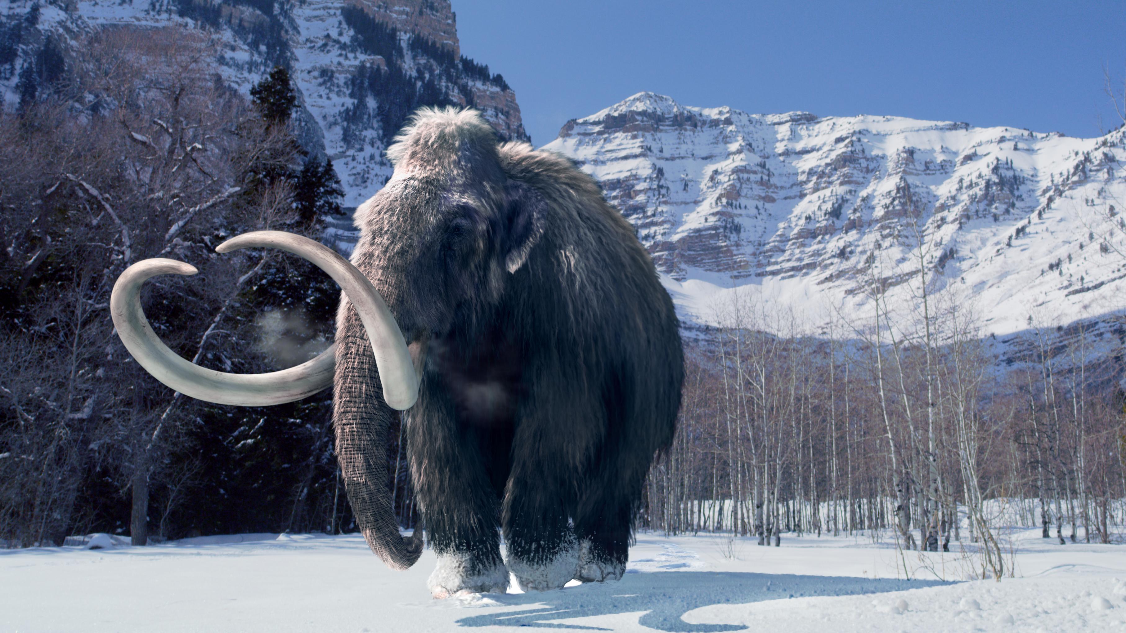
chemist John Asara of Harvard University, I obtained proteins form mammoth
fossils that were estimated to be 300,000 to 600,000 years old. Sequencing of
the proteins using a technique called mass spectrometry indentified them
unambiguously as collagen, a key component of bone, tendons, skin and other
tissues. The publication of our mammoth results in 2002 did not trigger much
controversy. Indeed, the scientific community largely ignored it. But our proof
of principle was about to come in very handy.
The next year a crew from the Museum of
the Rockies finally finished excavating another T. rex skeleton, which
at 68 million years old is the oldest one to date. Like the younger T. rex,
this one—called MOR 1125 and nicknamed “Brex,” after discoverer Bob Harmon—was
recovered from the Hell Creek Formation in eastern Montana. The site is
isolated and remote, with no access for vehicles, so a helicopter ferried
plaster jackets containing excavated bones from the site to the camp. The
jacket containing the leg bones was too heavy for the helicopter to lift. To
retrieve them, then, the team broke the jacket, separated the bones and
rejacketed them. But the bones are very fragile, and when the original jacket
was opened, many fragments of bone fell out. These were boxed up for me.
Because my original T. rex studies were controversial, I was eager to
repeat the work on a second T. rex. The new find presented the perfect
opportunity.
As soon as I laid eyes on the first piece
of bone I removed from that box, a fragment of thighbone, I knew the skeleton
was special. Lining the internal surface of this fragment was a thin, distinct
layer of a type of bone that had never been found in dinosaurs. This layer was
very fibrous, filled with blood vessel channels, and completely different in
color and texture from the cortical bone that constitutes most of the skeleton.
“Oh, my gosh, it’s a girl—and it’s pregnant!” I exclaimed to my assistant,
Jennifer Wittmeyer, She looked at me like I had lost my mind. But having
studied bird physiology, I was nearly sure that this distinctive feature was
medullary bone, a special tissue that appears for only a limited time (often
for just about two weeks), when birds are in lay, and that exists to provide an
easy source of calcium to fortify the eggshells.
One of the characteristics that sets
medullary bone apart from other bone types is the random orientation of its
collagen fibers, a characteristic that indicates very rapid formation. (This
same organization occurs in the first bone laid down when you have a
fracture—that is why you feel a lump in healing bone.) The bones of a
modern-day bird and all other animals can be demineralized using mild acids to
reveal the telltale arrangement of the collagen fibers. Wittmeyer and I decided
to try to remove the minerals. If this was medullary bone and if collagen was
present, eliminating the minerals should leave behind randomly oriented fibers.
As the minerals were removed, they left a flexible and fibrous clump of tissue.
I could not believe what we were seeing. I asked Wittmeyer to repeat the
experiment multiple times. And each time we placed the distinctive layer of
bone in the mild acid solution, fibrous stretchy material remained—just as it
does when medullary bone in birds is treated in the same way.
Furthermore, when we then dissolved
pieces of the denser, more common cortical bone, we obtained more soft tissue.
Hollow, transparent, flexible, branching tubes emerged from the dissolving
matrix—and they looked exactly like blood vessels. Suspended inside the vessels
were either small, round red structures or amorphous accumulations of red
material. Additional demineralization experiments revealed distinctive-looking
bone cells called osteocytes that secrete the collagen and other components
that make up the organic part of bone. The whole dinosaur seemed to preserve
material never seen before in dinosaur bone.
When we published our observations in Science
in 2005, reporting the presence of what looked to be collagen, blood vessels
and bone cells, the paper garnered a lot of attention, but the scientific
community adopted a wait-and see attitude. We claimed only that the material we
found resembled these modern components—not that they were one and the same.
After millions of years, buried in sediments and exposed to geochemical conditions
that varied over time, what was preserved in these bones might bear little
chemical resemblance to what was there when the dinosaur was alive. The real
value of these materials could be determined only if their composition could be
discerned. Our work had just begun.
Using all the techniques honed while
studying Big Mike, Rathonavis, Shuvuuia and the mammoth, I began an
in-depth analysis of this T.rex’s bone in collaboration with Asara,
who had refined the purification and sequencing methods we used in the mammoth
study and was ready to try sequencing the dinosaur’s much older proteins. This
was a much harder exercise, because the concentration of organics in the
dinosaur was orders of magnitude less than in the much younger mammoth and
because the proteins were very degraded. Nevertheless, we were eventually able
to sequence them. And, gratifyingly, when our colleague Chris Organ of Harvard
compared the T.rex sequences with those of a multitude of other
organisms, he found that they grouped most closely with birds, followed by
crocodiles—the two groups that are the closest living relatives of dinosaurs.
CONTROVERSY AND ITS AFTERMATH
Our papers detailing the sequencing work,
published in 2007 and 2008, generated a firestorm of controversy, most of which
focused on our interpretations of the sequencing (mass spectrometry) data. Some
dissenters charged that we had not produced enough sequences to make our case;
others argued that the structures we interpreted as primeval soft tissues were
actually biofilm—“slime” produced by microbes that had invaded the fossilized
bone. There were other criticisms, too. I had mixed feelings about their
feedback. On one hand, scientists are paid to be skeptical and to examine
remarkable claims with rigor. On the other hand, science operates on the
principle of parsimony—the simplest explanation for all the data is assumed to
be the correct one. And we had supported our hypothesis with multiple lines of
evidence
Still, I knew that a single gee-whiz
discovery does not have any long-term meaning to science. We had to sequence
proteins form other dinosaur finds. When a volunteer accompanying us on a
summer expedition found bones from and 80-million-year-old plant-eating
duckbill dinosaur called Brachylophosaurus canadensis, or
“Brachy,” we suspected the duckbill might be a good source of ancient proteins
even before we got its bones out of the ground. Hoping that is might contain
organics, we did everything we could to free it from the surrounding sandstone
quickly while minimizing its exposure to the elements. Air pollutants, humidity
fluctuations and the like would be very harmful to fragile molecules, and the
longer the bone was exposed, the more likely contamination and degradation
would occur.
Perhaps because of this extra care—and
prompt analyses—both the chemistry and the morphology of this second dinosaur
were less altered than Brex’s. As we had hoped, we found cells embedded in a
matrix of white collagen fibers in the animal’s bone. The cells exhibited long,
thin, branchlike extensions that are characteristic of osteocytes, which we
could trace from the cell body to where they connected to other cells. A few of
them even contained what appeared to be internal structures, including possible
nuclei.
Furthermore, extracts of the duckbill’s
bone reacted with antibodies that target collagen and other proteins that
bacteria do not manufacture, refuting the suggestion that our soft-tissue
structures were merely biofilms. In addition, the protein sequences we obtained
from the bone most closely resembled those of modern birds, just as Brex’s did.
And we sent samples of the duckbill’s bone to several different labs for
independent testing, all of which confirmed our results. After we reported
these findings in Science in 2009, I heard no complaints.
Our work does not stop here. There is
still so much about ancient soft tissues that we do not understand. Why are
these materials preserved when all our models say they should be degraded? How
does fossilization really occur? How much can we learn about animals from
preserved fragments of molecules? The sequencing work hints that analyses of
this material might eventually help to sort out how extinct species are
related—once we and others build up bigger libraries of ancient sequences, and
sequences from living species, for comparison, As these databases expand, we
may be able to compare sequences to see how member of lineage changed at the
molecular level. And by rooting these sequences in time, we might be able to
better understand the rate of this evolution. Such insights will help
scientists to piece together how dinosaurs and other extinct creatures
responded to major environmental changes, how they recovered from catastrophic
events, and ultimately what did them in.
Comments
+52011-08-11
10:19
Ms. Mary
Schweitzer asks, "Why are these materials preserved when all our models
say they should be degraded?" A simple $600 experiment test for C-14 in
one of 10 Accelerated Mass Spectrometer (AMS) laboratories in the USA would
answer that. The half life for the radioactive decay of C-14 is 5,730 years and
AMS equipment can detect each atom of C-14 with reasonable accuracy to about
eight half-lives or about 50,000 years.
Since 1990
there has been a steady stream of reports of finding C-14 in dinosaur bones and
other “ancient” fossils with a definitive report being published in a book
written in 2009 entitled "Evolutionism: The Decline of an
Hypothesis." C-14 dates of 23,170 ±170 to 30,890 ± 200 years were reported
for dinosaur bone collagen in the paper entitled: “Recent C-14 Dating of
Fossils Including Dinosaur Bone Collagen. The results appear to be a
confirmation of rapid formation of the geologic column as modern sedimentology
studies have predicted.”
....


